Unraveling the inner workings of cells with labels
17 October 2024

Cells are the fundamental building blocks of life. Cell biology, the study of cell structure, function and interactions, is therefore essential for nearly all aspects of modern biology and medicine. For instance, cell research helps us understand and treat diseases such as cancer and diabetes. Additionally, cell biology can play a role in agriculture, such as growing better crops by genetically modifying them.
Technological advances have drastically changed cell research in recent decades. Advanced microscopes make it possible to study cells and their molecular processes in detail at an unprecedented level. A challenge here, however, is that both the cells and their components and all substances in them are transparent. 'In order to still be able to see something in those cells, we make special dyes,' says Joachim Goedhart, assistent professor Molecular Cytology at the Swammerdam Institute for Life Sciences (SILS) of the University of Amsterdam.
Fluorescent labels
What's special about the dyes that Goedhart and his colleagues are developing is that you don’t simply add them to the cells under the microscope like a kind of paint. Instead, they design pieces of DNA code that they introduce into the cells, after which the cells themselves produce the desired dyes via that DNA. These labels are also fluorescent: they emit colored light when viewed under special fluorescence microscopes.
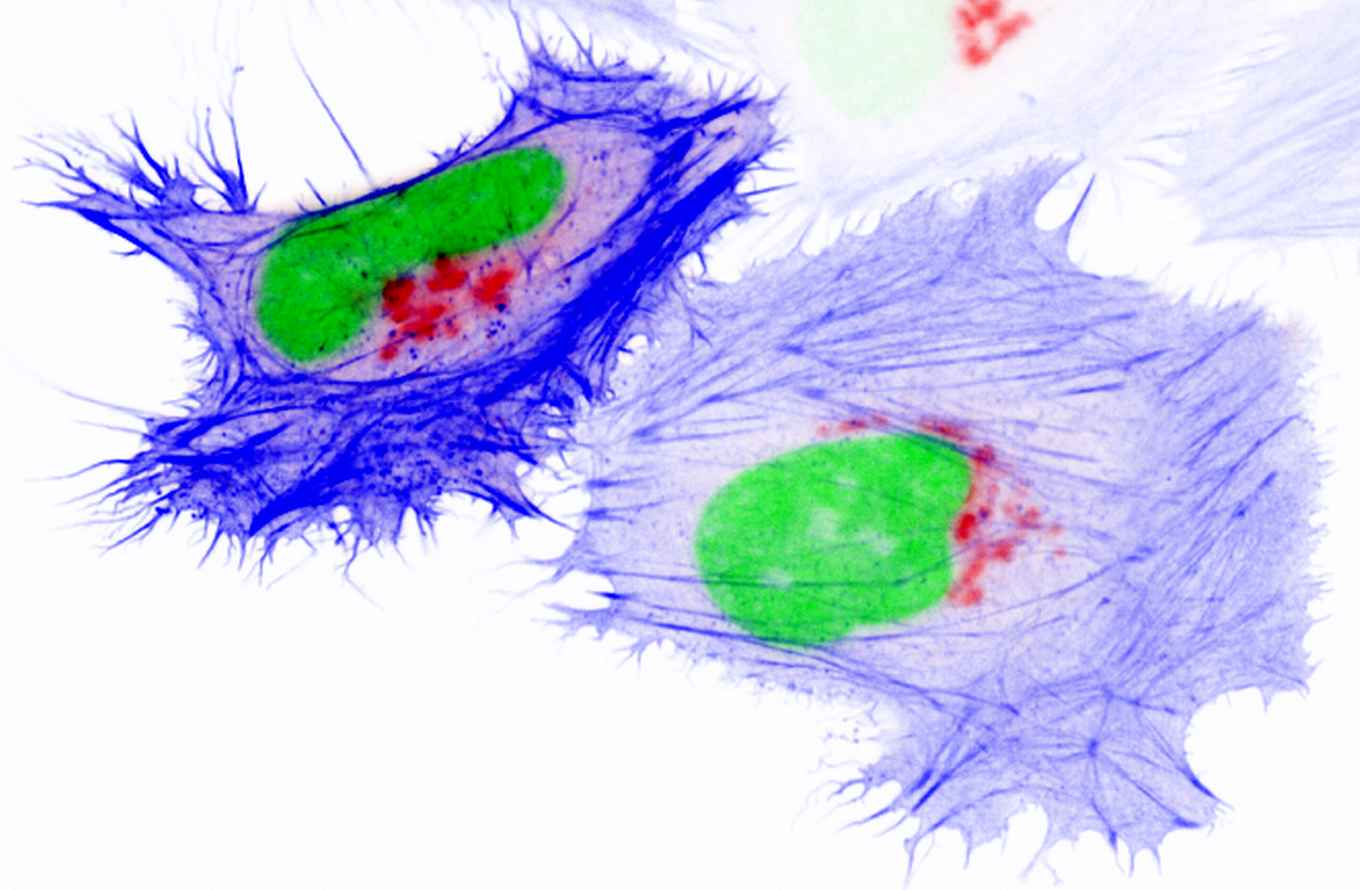
Using this colored light, cell biologists can essentially tag parts of cells or substances within it. Goedhart distinguishes two types of probes. The "dumb" propes only make parts of the cell visible. The "smart" probes can also report what is happening in the cell and detect the presence of specific molecules.
Tinkering with DNA
Both types of probes are used extensively, for example for labelling cancer cells, tracking viral infections in cells, and analysing neural networks in the brain. However, the probes don't work equally well for every cell type, and they emit less and less light over time.
That’s why the Molecular Cytology research group focuses on optimising these probes. They do this by modifying the DNA code of the probes, and creating many variants. Ultimately, they select the best probe for specific applications. For this, they use their own advanced microscopes from the Van Leeuwenhoek Centre for Advanced Microscopy (LCAM)-FNWI.
When optimising the probes, the researchers evaluate various properties. Goedhart: ‘With the dumb probes, you especially want the label to be very clear. We also want them to continue to emit light stably, because we follow them for a long time.’ In addition, the probes shouldn't damage the cell. Smart probes must also be able to report well on a specific cellular process.
Very accurate sensors
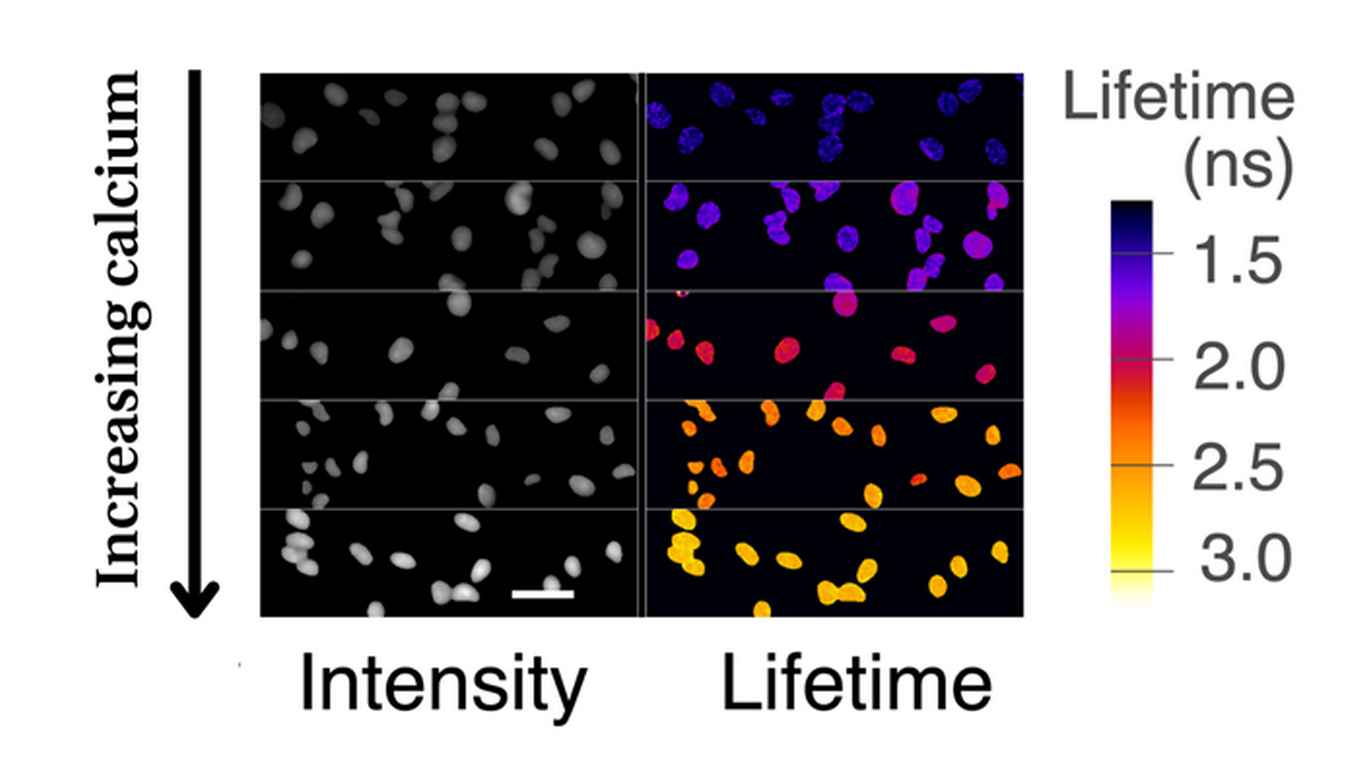
A calcium probe is an example of a smart label, which allows researchers to measure the calcium concentration in a cell. The light intensity of the probe depends on the calcium concentration in the cell. It's therefore important that changes in intensity can be measured by scientists. However, it's difficult to measure the exact calcium concentration, especially because the light intensity depends on external factors, such as the microscope settings.
For applications in cell biology, it is very important to know the exact concentration. To address this, Goedhart's research group has developed a new probe. Scientists can use this to measure the calcium concentration based on the probe's lifespan. Goedhart: 'This new probe provides an absolute measurement. This means that the measurement is exactly the same across different devices. We think this is a fantastic new measuring method.'
Controlling cells
Goedhart is also very enthusiastic about his work with light-sensitive proteins in cells. These proteins originally come from plants and can stick together under the influence of light. They can be used to control certain cell processes very precisely with light. Neurologists use this technology to easily activate or deactivate neurons.
Goedhart's research group has optimized these light-sensitive proteins for research into blood vessel walls, in collaboration with Professor Jaap van Buul of the Amsterdam UMC. Goedhart: 'Certain cells in your blood vessels form a barrier so that your blood doesn't enter your body. We have been able to use this technology to make the cells connect better to each other, so that they form a better barrier. In this way. you can use light to control the shape and behaviour of cells. It’s truly amazing that this is possible.’