NL-BioImaging receives 15 million euros from the NWO National Roadmap programme
21 February 2023
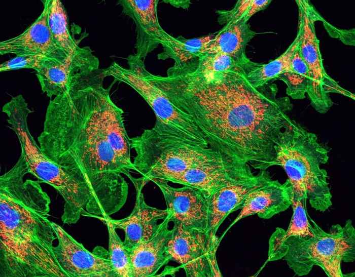
NL-BioImaging is the Dutch scientific infrastructure for advanced microscopy. It stands on the shoulders of famous Dutchman Antoni van Leeuwenhoek who studied 'tiny animals' and processes such as reproduction with his own microscope. Now, 300 years after his death, turbulent developments in microscopy make it possible to visualize processes in living cells, organoids and organisms with newly developed biosensors. The revolutionary insights thus obtained lead to scientific breakthroughs and advanced applications in society, such as a better understanding of diseases like cancer, metabolic diseases, cardiovascular diseases and brain disorders and the development of new strategies for their prevention, therapy and cure.
The Consortium
NL-BioImaging is a collaboration of and by all Dutch universities, UMCs and other knowledge institutions such as the Netherlands Cancer Institute. All the researchers involved work together for optimal innovation, integration and data analysis. The ambitions are too great to be realized by any individual institution and thus require a joint approach, which is made possible by the currently obtained funding (25 million euros, of which 15 million by NWO).
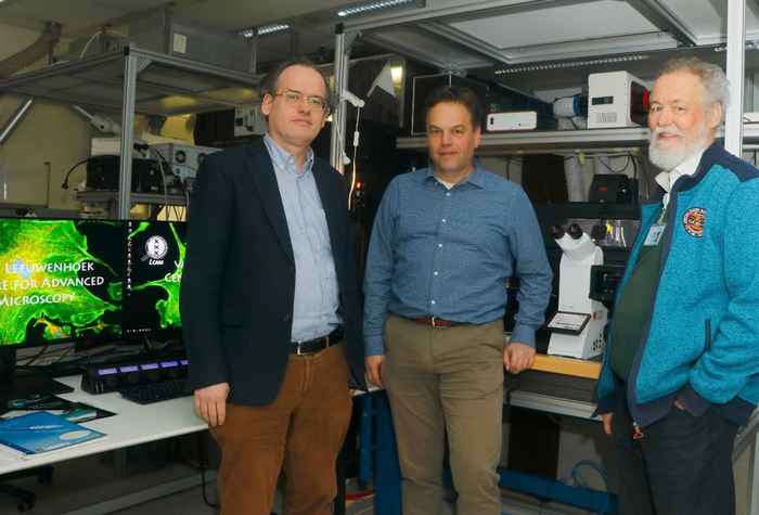
The Amsterdam UMC and the UvA coordinate the consortium, with a leading role for the Van Leeuwenhoek Center for Advanced Microscopy (LCAM) in which researchers from Amsterdam UMC, the UvA’s Faculty of Science and the Netherlands Cancer Institute (NKI) work together. Since its establishment in 2009, LCAM has played a pioneering role in organizing the Dutch microscopy landscape and placing it on the National Roadmap for large-scale research infrastructure. After NL-BioImaging was placed on the National Roadmap in 2012, this Dutch research infrastructure also became part of the Euro-BioImaging ESFRI large-scale research infrastructure formalized in 2019. Within Euro-BioImaging, LCAM has been ratified as a Functional Imaging Flagship node with a focus on visualizing and quantifying processes in living cells such as protein aggregation (Huntington's disease), signaling (developmental biology and cancer) and blood cell mobility (cardiology and immunology).
New state-of-the-art equipment
With the financing of the NL-BioImaging Roadmap, all participating Dutch microscopy centres can bring their infrastructure to the top level, specific techniques and expertise are connected, and a national network for data analysis and management will be set up. This opens the door to new scientific breakthroughs by thousands of life scientists in the Netherlands. In LCAM Amsterdam, for example, 4 million will be invested in equipment and 1.5 million in support, which gives an enormous boost to state-of-the-art microscopy cell biology. At the University of Amsterdam, under supervision of Dorus Gadella (LCAM-UvA and Swammerdam Institute for Life Sciences), investments will be made in a new super-resolution microscope, which produces 3-5 times sharper images than conventional microscopes by making smart use of shortening of the (nanosecond 10-9 s) fluorescence lifetime. Gadella: ‘We were able to test this concept with new switchable fluorescent proteins and biosensors developed by ourselves, so we know this technique will actually work in living cells and yield dynamic super-resolution information.’
Consortium leader Eric Reits (LCAM – Amsterdam UMC) will install a high-content 3D microscope with which fast processes with fluorescent biosensors in organoid models can be analysed on-the-fly. Reits: ‘Using self-steering microscopy scripts, the microscope is fast enough to quantify the effects of potential therapeutic strategies and to isolate cells with an interesting phenotype for proteome, metabolome and genome analysis, i.e. visual omics.’ Kees Jalink (LCAM-NKI) invests in the development of automated pooled microscopy screening, in which extensive compounds and CRISPR knockout libraries are tested in cells for changes in phenotype. Jalink: ‘By using different fluorescent biosensors that we develop at LCAM, researchers can screen which compounds and genes effectively change a phenotype in cells.’
Education
All these developments put LCAM and Amsterdam further on the map. Since Gadella and Reits coordinate the unique biomedical Master's track Cell Biology and Advanced Microscopy, in collaboration with Jalink, the new technologies and analysis scripts will also be used immediately in education, thus training the new generation of cell biologists. Reits: 'Within this master's program, students learn hands-on how to make optimal use of the various microscopes and they learn about the possibilities of AI data analysis. Combined with internships at (inter)national microscopy expertise groups, they are well prepared to immediately implement these state-of-the-art techniques in biomedical research as PhD students.'