UvA biologists develop a new fluorescent biosensor to measure calcium levels in cells
10 December 2021
The revolution of using fluorescent proteins to stain parts of cells began with the discovery of how the now-famous Green Fluorescent Protein (GFP) produced by the jellyfish Aequoreae victoria can be used in the lab. The UvA’s laboratory of Molecular Cytology, part of the Swammerdam Institute for Life Sciences, has been developing and improving stains and biosensors of different colors that are inspired by GFP for several years. By altering the genetic code of the GFP protein bright variants of for instance cyan and red fluorescent proteins are created, with adjustable properties. The code and the DNA can be shared with other labs and enables scientists to use these stains and biosensors for their own research.
Calcium
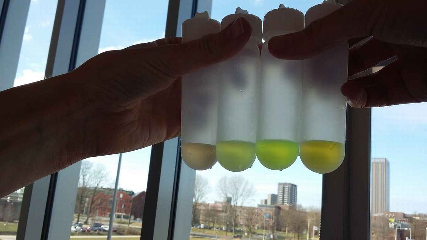
As part of her PhD project, Franka van der Linden took on the quest to develop a cyan-colored fluorescent protein that can be used as a biosensor to visualize calcium levels in cells. Calcium ions play a key role in a large number of cellular processes. They are for example needed for the contraction of muscles, they are involved in brain activity, and calcium ions are released after the perception of hormones by cells. Given the universal role of calcium in cellular processes, it is no wonder scientists are interested in measuring calcium concentrations.
Challenges
Joachim Goedhart, senior author of the paper in Nature Communications that elaborates on the new fluorescent protein, explains some of the challenges the team encountered: ‘The standard approach for measuring changes in concentration of for example calcium within cells is to simply measure the amount of light that the biosensor emits with a microscope. But, the amount of emitted light also depends on the concentration of the added biosensor. This makes it very difficult to accurately quantify calcium concentrations solely based on the amount of emitted light. What we did was take a slightly different approach: we used the so-called fluorescence lifetime as a measure. Fluorescence lifetime does not depend on the fluorescence intensity and is therefore an ideal parameter for measurements in cells. It can be measured with a dedicated microscope, a Fluorescence Lifetime Imaging Microscope or FLIM.’
It took the team many months of making and trying different, slightly modified DNA codes. But eventually they managed to create a protein that shows a fluorescent lifetime change when calcium concentrations change. Goedhart: ‘A very nice side-effect of using this cyan fluorescent protein is that it’s not sensitive to acid. Whereas many GFP-based fluorescent proteins lose their fluorescence when the pH drops, the cyan one is not affected by pH changes.’
Tested in practice
Van der Linden, Goedhart and their colleagues have also already tested the use of the new fluorescent protein in practice. Goedhart: ‘We used it to detect the increase of the calcium concentration in endothelial cells by stimulation with histamine. Additionally, in collaboration with Jaap van Buul of Sanquin Research, we used the protein to detect calcium concentrations in a layer of endothelial cells, mimicking the inner layer of blood vessels. Another demonstration was done in collaboration with Joep Beumer and Hans Clevers of the Hubrecht Institute, Utrecht. In their lab, an organoid model of the human gut was engineered to express the biosensor. The mini-guts were brought to Amsterdam where the lifetime imaging microscope was used to measure calcium. The calcium dynamics were precisely quantified, which shows the potential of our biosensor as a new and accurate measurement device.’
Details of the publication
Franka H. van der Linden et al, A turquoise fluorescence lifetime-based biosensor for quantitative imaging of intracellular calcium, in: Nature Communications, 10 November 2021, DOI: https://doi.org/10.1038/s41467-021-27249-w